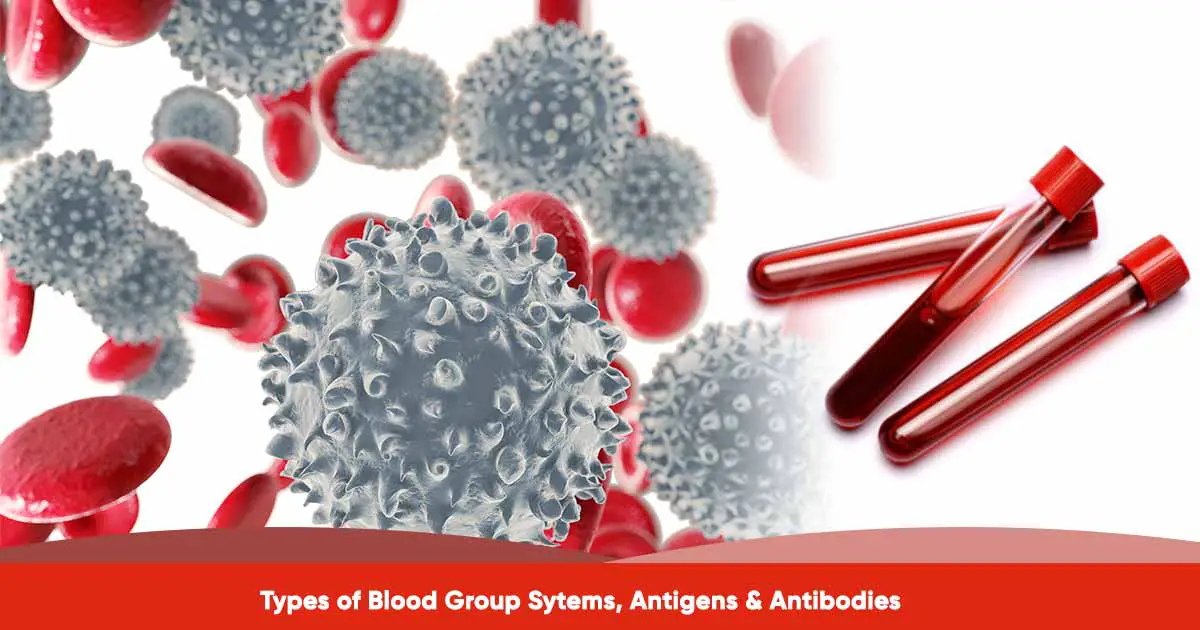
Blood grouping or blood typing is the grouping of blood based on the presence or absence of antigens on the surface of the red blood cells. These antigens may be proteins, carbohydrates, glycoproteins, or glycolipids.
International Society of Blood Transfusion (ISBT) recognizes 43 blood group system, with over 349 antigens. 48 genes genetically determine these 43 systems. According to ISBT, three categories of antigens are not yet linked to any blood group system.
The most important blood group systems are ABO and Rh. Before we explain the different blood group types, we will explain antigens and antibodies.
Blood Group Antigens
Antigens are parts of the membrane structure. They are a complex structure with proteins, and carbohydrates and offer structural support. Antigens are produced when one inherits specific genes. Also, the genes produces different antigen options within one blood group system.
The red cells have blood group antigens, while white cells and platelets have HLA antigens. Platelets also contain additional HPA antigens. In blood donation, reaction occurs when the antigen of the donor cells react with the antibody of the patients’ plasma.
During transfusion, the antigens on the red cells of the donor will stimulate antibody production, especially if the patient lacks the antigen.
Antigens are also called agglutinogens because they have the ability to cause agglutination of RBCs.
Blood Group Antibodies
Blood group antibodies are protein molecules called immunoglobulins (Ig). Antibodies are produced by the immune system in response to antigen and are found in the serum/plasma. The production are stimulated during blood transfusion, environmental factors (ie naturally acquired as with anti-A and anti-B), or during pregnancy or childbirth. The fetal antigen enters maternal circulation, stimulating antibody production in the mother.
In the red cells, they bind to the corresponding antigen, leading to destruction of the red cells. In the body, antibody-antigen reactions destroys the cells directly when cells break up in the bloodstream (intravascular). It can be indirectly when organs in the body such as the liver and spleen removes the antibody coated cells (extravascular).
In the lab, the antibody-antigen reaction result in agglutination, the clumping of red cells into visible agglutination. This is caused by antibody and antigen crosslinking.
Agglutination in the lab can help to detect incompatible match, red cell antigens (‘blood grouping’), and antibodies in the plasma.
Different Blood Grouping System
As said earlier, there are 36 blood grouping system. But some selected blood group systems are:
| Blood Group System | Symbol | Gene name | No of antigens |
| ABO | ABO | ABO | 4 |
| Rh | RH | RHD, RHCE | 56 |
| MNS | MNS | GYPA, GYPB, (GYPE) | 50 |
| Lutheran | LU | BCAM | 26 |
| Duffy | FY | ACKR1 | 5 |
| Kidd | JK | SLC14A1 | 3 |
| Kell | KEL | KEL | 3 |
| Lewis | LE | FUT3 | 6 |
| P1PK | P1PK | A4GALT | 3 |
| Landsteiner – Wiener | LW | ICAM4 | 4 |
| Diego | DI | SLC4A1 | 23 |
ABO Blood Group
The ABO group is the most important blood group system in human blood transfusion. The system is also available in other animals like rodents, apes, chimpanzees, gorillas, and bonobos. Immunological reaction between the antigen and antibody determines the ABO group systems.
Karl Landsteiner discovered the ABO systems in 1901, and later received the Nobel Prize in 1930 for his work in Medicine. In 1902, two scientists, Adriano Sturli and Alfred von Decastello, who worked under Landsteiner, discovered the type AB.
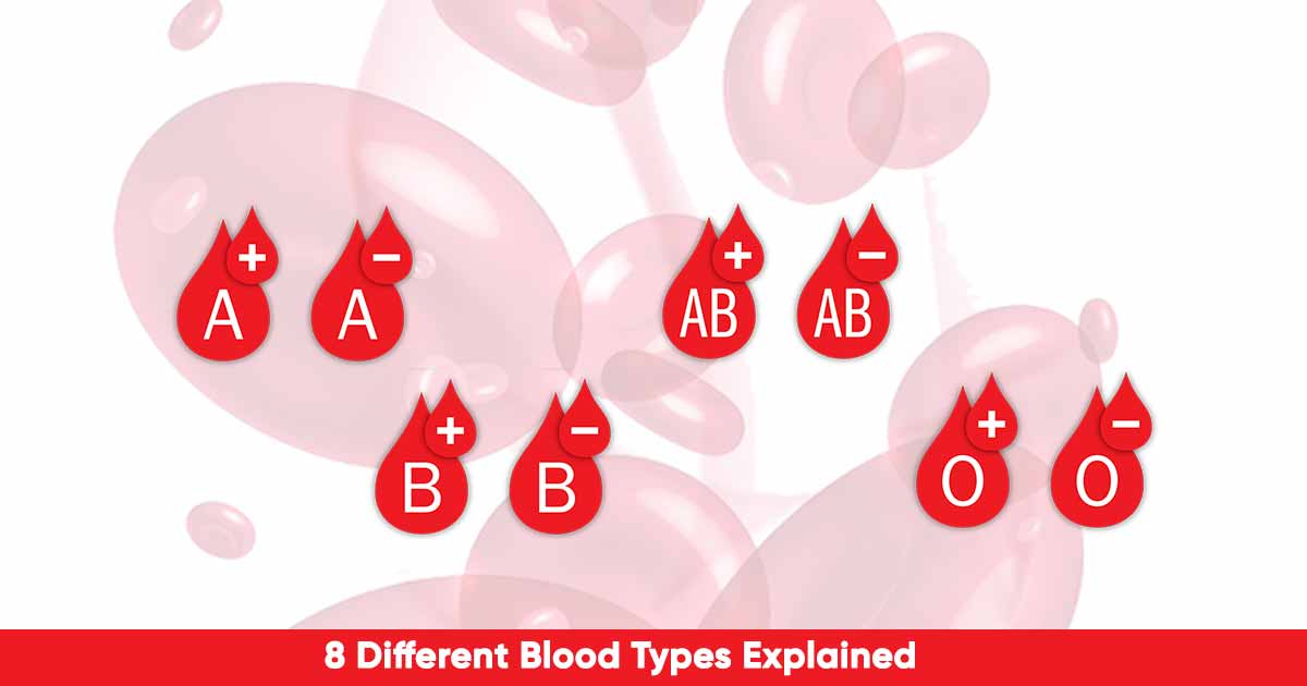
In 1907, Jansky classified the blood groups into four – A, B, AB, O, and this system is still in use today. There is a further classification into 8 blood types during transfusion.
Though there have been earlier records of blood transfusion, Reuben Ottenberg was the first to record the pre-transfusion blood testing result to ensure compactibility. He transfused the blood between two persons at Mount Sinai Hospital in New York.
Landsteiner Rule: According to the Landsteiner Rule, If an antigen is present on a patient’s red blood cells (RBCs) the corresponding antibody will NOT be present in the patient’s plasma, under ‘normal conditions’.
Understanding the ABO Blood Group System
The ABO blood group is done using the agglutination principle. Agglutination is the coming of separate particles, such as the red blood cells, into clumps, or masses. It occurs when the antigen mixes with antibody, which is called isoagglutinin. For example, mixing A antigen with anti-A, or B antigen with anti-B antibody.
The ABO blood group is divided into ‘A, B, AB and ‘O’ group based on the presence or absence of antigen A and antigen B.
- ‘A’ group: The blood has antigen A, and β-antibody in the serum.
- ‘B’ group: Blood contains antigen B and α-antibody. This group is found in 35% of Asian population.
- ‘AB’ group: This group has both antigen A and B, but has no antibody.
- ‘O’ group: This group has no antigen, but contains both α and β antibodies in the serum. Some African tribes are 100% ‘O’ group.
In blood transfusion, the antigen of the person receiving the blood should match with the antibody of the person donating the blood.
The ‘O’ group (universal donor)
The ‘O’ group has no antigen on their red blood cells, and so there will be no agglutination. This means they can give blood to any other group. They are called universal donors.
The AB group (universal recipients)
The AB group has no antibody in their plasma. This means there is no agglutination from other blood groups. They can receive blood from any group, hence they are called universal recipients.
In mismatched blood transfusion, there is reaction between the plasma of the recipient and the red blood cells of the donor. If the donor’s plasma has agglutinins against the red blood cells of the recipient, there will be no agglutination as the antibodies are diluted in the recipient blood.
Transfusion reaction can be as mild as fever, and hives to severe reactions such as renal failure, shock, and death.
| ABO Group | Antigen Present | Antigen Missing | Antibody Present |
| A | A | B | Anti-B |
| B | B | A | Anti-A |
| O | None | A and B | Anti-A and B |
| AB | A and B | None | None |
How do I inherit the blood type?
Using a table, let us illustrate how blood types are inherited.
| Father | Mother | A | B | AB | O |
| O | O | ✅ | |||
| O | A | ✅ | ✅ | ||
| O | B | ✅ | ✅ | ||
| A | A | ✅ | ✅ | ||
| A | B | ✅ | ✅ | ✅ | ✅ |
| B | B | ✅ | ✅ | ||
| AB | O | ✅ | ✅ | ||
| AB | A | ✅ | ✅ | ✅ | |
| AB | B | ✅ | ✅ | ✅ | |
| AB | AB | ✅ | ✅ | ✅ |
The Rh Blood Group System
The Rh blood group system is the second most important blood grouping system after the ABO system.
The system consists of 50 defined blood-group antigens, with six common types of Rh antigens. Each of the Rh antigen is called Rh factor, and designated C, D, E, c, d, and e.
Of all the Rh antigen types, the most antigenic and predominant in the population is the type D antigen. Anybody with type D antigen is called Rh positive, or D positive, while those without the antigen are called Rh negative, or D negative. 80% of D negative people produce anti-D when exposed to the D antigen.
RhD-negative blood is distributed in 14% of Caucasians, 5—10% in black, and 0.3% of Chinese people.
In 1939, Philip Levine and Rufus Stetson used an immunized pregnant woman to describe the importance of the rhesus system. She suffered adverse reaction after receiving a blood transfusion from her husband. This transfusion was done after she gave birth to a still-born.
However, the Rh blood group system was discovered by Karl Landsteiner and A.S. Weiner in 1940. It was discovered in Rhesus macaque, a species of monkey, hence the name, ‘Rh factor’.
The immune reaction suffered by the woman as reported by Levine and Stetson was called “erythroblastosis fetalis”. It describes the fetal symptoms that happened as a result of the exposure of the mother to antigen not present in her red blood cells but in the father, and also inherited by the fetus. The antigens were later called the “Rh factor”, D antigen, or RhD.
Erythroblastosis Fetalis or Hemolytic disease of the fetus and newborn (HDFN)
Erythroblastosis Fetalis or Hemolytic Disease of the Fetus and Newborn (HDFN), occurs due to incompatibility between the mother and child blood. HDFN, is mostly called “Rh Disease”. When an expectant mother is RhD-negative, and the fetus is RhD-positive, (after inheriting the RhD antigen from the father), the mother will be exposed to RhD antigen on the fetal red blood cells during childbirth.
This will prompt the immune system to make antibodies in the mother, leading to the development of anti-Rh agglutinins. These agglutinins diffuse from the placenta into the fetus, causing red blood cell agglutination in the fetus.
It rarely affects the first pregnancy, but subsequent pregnancies, due to the presence of the antibody in the mother. Hence, there is significant risk of HDFN if the fetus is RhD-positive.
Signs of HDFN in a fetus are enlarged liver spleen, heart fluid buildup in the fetus’ abdomen when observed through ultrasound. In a newborn, there are signs and symptoms such as jaundice, enlarged liver and spleen, severe edema, dyspnea (difficulty in breathing) and anemia causing pale appearance in a baby.
According to the American College of Obstetricians and Gynecologists (ACOG), one out of seven fetuses affected by the RhD HDFN suffers mortality if preventive measures are not taken.
The recommendation of ACOG is for all RhD-negative expectant mother to receive RhD immune globulin (RhIG) injection around the 28th week of pregnancy, and shortly after the delivery of RhD-positive newborn. This prevents RhD alloimmunization, with a success rate of 98.4 to 99%.
Other Less Common Blood Grouping System
Kell Blood Group System
They are the third most important immunogenic antigen after the ABO system, and Rh system. Since the first discovery, there are now 25 Kell antigens. The antibody is anti-K. This anti-K antibody causes severe hemolytic disease of the fetus and newborn (HDFN) and haemolytic transfusion reactions (HTR).
This blood group system was first discovered in the serum of Mrs. Kellacher, who suffered hemolytic reaction after reacting to the erythrocytes of her newborn infant.
Duffy System
The Duffy-antigen was first isolated in a hemophilia patient, Duffy. Also known as Fy glycoprotein, it is in the surface of the red blood cells. It is a nonspecific receptor for several chemokines and also acts as a receptor for human malarial parasite, Plamodium vivax.
Antigens Fya and Fyb on the Duffy glycoprotein can generate four possible phenotypes – Fy(a+b−), Fy(a+b+), Fy(a−b+), and Fy(a−b−). The antibodies are IgG subtypes, and can cause hemolytic transfusion reaction.
MNS Antigen System
This system is based on two genes – Glycophorin A and Glycophorin B. This was first described by Landsteiner and Levine in 1927. The blood group is under the control of an autosomal locus on chromosome 4, and a pair of co-dominant alleles LM and LN.
The antibodies – Anti-M and anti-N antibodies are mostly IgM types, and rarely do blood transfusion reaction occurs.
Kidd Blood Group System (Jk antigen)
Kidd antigen is a glycoprotein on the membrane of red blood cell surface acting as urea transporter for the red blood cells and renal endothelial cells. The Kidd antibodies are rare, but cause transfusion reactions that can be severe. The antigen react with antibody, anti-Jka. This was first observed in Mrs Kidd, who delivered a baby with hemolytic disease of the fetus and newborn (HDFN).
Kidd blood group system has antigen Jka, the first discovered antigen, and two others – Jkb and Jk3.
Lutheran system
In this system, there are four pairs of allelic antigens. This represents a single amino acid substitution in the Lutheran glycoprotein at chromosome 19. Rare antibody reaction against this blood group type.
References:
- https://www.isbtweb.org/isbt-working-parties/rcibgt.html
- https://www.isbtweb.org/resource/tableofbloodgroupsystems.html
- https://www.researchgate.net/publication/270005501_Blood_groups_systems
- https://www.gputtawar.edu.in/downloads/blood%20group.pdf
- https://www.bloodgroupgenomics.org/blog-blood-experts/articles/history-rh-blood-group-how-genomics-changing-rhd-typing/

